Neurophysiological correlates of artistic image creation by representatives of artistic professions
Abstract
The steadily increasing demand for artistic professions brings to the fore the task of studying the phenomenon of art by researching the unique capacity of the human brain to create works of art in different spheres of creative activity. So far, only a few studies have investigated creativity-related brain activity in representatives of the creative professions. The aim of the empirical research was to study the neurophysiological correlates of artistic image creation by representatives of the artistic professions. The participants were 60 right-handed females aged 23-27, divided into three groups— artists (23 people), actors (17 people), and specialists who do not work in an artistic field (20 people). The mono-typing technique was used to model the creative artistic process. EEG signals were recorded in a resting state, and during four stages of the creation of an artistic image (viewing of monotypes, frustration, image creation, and thinking over the details) from 21 electrodes set on the scalp according to the International 10-20 System. We analyzed EEG coherence for each functional trial at theta (4.00–8.00 Hz), alpha1 (8.00–10.5 Hz), alpha2 (10.5–13.00 Hz), and beta (13.00– 35.00 Hz) frequency bands. For statistical analysis, we used MANOVA and post hoc analysis. We found that the neurophysiological correlates of creating an artistic image are different at different stages of the creative process, and have different features for artists and actors. The actors primarily show dominance of right hemisphere activity, while close interaction of the hemispheres distinguishes the brains of the artists. The differences revealed in brain cortex functioning when artists or actors create an artistic image reflect different strategies of imaginative creative work by representatives of these professions.
Received: 13.04.2016
Accepted: 30.07.2016
Themes: Neuropsychology; 120th anniversary of Lev Vygotsky
PDF: http://psychologyinrussia.com/volumes/pdf/2016_4/psychology_2016_4_5.pdf
Pages: 51-72
DOI: 10.11621/pir.2016.0405
Keywords: brain hemispheres, cortex, EEG, coherence, frequency band, art, artistic image
Introduction
Art is an essential part of human life, a specific area of human activity aimed at cognition of objective reality. Artistic imagery is a way of exploring reality. An artistic image is a merging of immediate data, sensual characteristics, and the idea that is used to convey the artist’s general attitude.
Artistic, engineering, musical, and dance activities, as well as performance, require the creation of an artistic image, represented by the different forms of art (picture, dance, piece of music). Each of these professional activities has its structural peculiarities, its means of creating and artistically realizing the valuable contents of the image, as well as its methods of teaching professionals. Hence, the artist’s professionalism is rooted in a special competence predetermining the level of mastery of composition based on the development of compositional thinking and practical graphic activity. On the other hand, the creative work of actors is expressed through forming scenic images, often employing improvisation that adds freshness and spontaneity to the performance.
The steadily increasing demand for the artistic professions brings to the fore the task of studying the phenomenon of art by researching the unique capacity of the human brain to create works of art in different spheres of creative activity. The neuroscientific study of creative activity helps us understand how the brain works during the generation of creative ideas. The study of the development of higher mental functions led Vygotsky (1934/2005) to the conclusion that the brain’s role in its own organization must change in each individual’s development. Taking into account that the creative process is a complex cognitive activity, we are guided by Vygotsky’s idea about the connection between the development of mental functions and change in the organization of the brain.
Understanding the brain’s mechanisms during creative processes will permit us to carry out psychophysiological diagnostics aimed at vocational guidance; forecast success in training artists; and select and implement programs of potential creative development.
Existing studies of the neurophysiological correlates of artistic creativity are promising. But so far, only a few studies have investigated creativity-related brain activity (like improvising music, composing an artwork) in artists (Bhattacharya & Petsche, 2005; Dikiy, Dikaja, & Karpova, 2016; Kowatari, Lee, Yamamura, Nagamori, Levy, Yamane, & Yamamoto, 2009), musicians (Dikaya & Skirtach, 2015; Dikiy, Dikaya, & Skirtach, 2014; Gibson, Folley, & Park, 2009; Limb & Braun, 2008; Liu, Chow, Xu, Erkkinen, Swett, Eagle, & Braun, 2012; Pereda, Rahman, & Bhattacharya, 2014; Pinho, de Manzano, Fransson, Eriksson, & Ullén, 2014; Villarreal , Cerquetti, Caruso, Aranguren, Gerschcovich, Frega, & Leiguarda, 2013), actors (Rodionov, 2013; Starchenko, Kireev, & Medvedev, 2014); and dancers (Dikaya, Naumova, & Naumov, 2015; Fink, Graif, & Neubauer, 2009).
However, the question of the neurophysiological correlates of professional creative activity has been left unanswered, despite the growth of scientific interest in studying the brain functioning of representatives of the artistic professions. Thus, research from recent years reveals the contradictory character of contemporary scientists’ ideas about the role of the brain’s hemispheres in connection with creative work. The results of some research state that the right hemisphere dominates in the process of creative activity (Asari, Konishi, Jimura, Chikazoe, Nakamura, & Miyashita, 2008; Bhattacharya & Petsche, 2005; Jung, Mead, Carrasco, & Flores, 2013; Jung-Beeman, Bowden, Haberman, Frymiare, Arambel-Liu, Greenblatt, & Kounios, 2004; Kounios & Beeman, 2014), while others show left hemisphere dominance (Dikiy, Dikaya, & Skirtach, 2014; Gonen-Yaacovi, de Souza, Levy, Urbanski, Josse, & Volle, 2013; Jin, Kwon, Jeong, Kwon, & Shin, 2006). The scientific literature also contains studies that show close inter-hemisphere integration (Jauљovec, 2000; Mayseless & Shamay-Tsoory, 2015; Razumnikova & Yashanina, 2014), and independent functioning of the brain hemispheres while the person is performing creative work (Bechtereva & Nagornova, 2007; Dikaya, Dikiy, Karpova, & Lavreshina, 2016; Fink & Benedek, 2014).
The likely answer concerning the cerebral hemispheres is that both are functional in creativity, but each hemisphere contributes a different facet, still little understood, to the creativity process (Zaidel, 2014).
Questions about the level of cortical activation when a person is performing creative tasks, and about the role of different regions of the brain in the implementation of creativity, remain open as well.
The question of the source of the original ideas in artistic work is a complex one that researchers would like understand. Altogether, the latest studies suggest that creativity cannot be localized in one or a few regions of the brain; rather they show that when humans are engaged in any sort of creative activity, a multitude of regions of the brain are activated (Dietrich & Kanso, 2010; Dikaya & Dikiy, 2015; Starchenko et al., 2014; Wiggins & Bhattacharya, 2014).
We can conclude that there is significant heterogeneity in the results of studies of brain functioning during creative work. The contradictory data received by researchers can be explained by the fact that studies of the brain’s organization during the creative process are carried out without regard to its basic stages. There is a deficiency of research aimed at studying the dynamics of brain activity at different stages of creative process, which are: 1) Preparation (defining a problem, gathering information, attempting a solution); 2) Incubation ( the unconscious process of the “maturing of the decision”); 3) the Illumination or Insight or “Aha” experience ( a creative idea flashes into sight); and 4) Verification (checking the adequacy of the solution).
A promising approach involves EEG research of the brain’s cortex’s functional organization in representatives of the different artistic professions on a task close to actual professional life. We believe that a mono-typing technique such as artistic improvisation based on a shapeless smudge can be a good way to model the real creative process.
Method
The aim, hypotheses, and participants
The aim of our empirical research was to study the neurophysiological correlates of artistic image creation by representatives of the artistic professions.
The following assumptions were set forward as hypotheses:
- The main stages of artistic image creation can be reflected in the dynamics of the brain activity of representatives of different artistic professions;
- The dynamics of brain activity at different stages of artistic image creation can be predetermined by the type of professional activity (art sphere) that the representatives are engaged in (artists, actors).
Sixty right-handed female human subjects aged 23-27 took part in the research. They were divided into three groups depending upon their profession: artists (23 people), actors (17 people), and specialists who do not work in an artistic field (20 people). All participants were right-handed at birth; none were “corrected” righthanders. Handedness was assessed using the Questionnaire of Hand Preference (Annett, 1970).
All the artistic professionals had higher or vocational education and 2-3 years of work experience in their professions. Taking these indicators into account provides the basis for assuming the subjects had a definite level of professional development in carrying out creative work, and for avoiding the possibility of professional deformation influencing the subjects’ personalities.
The participants were briefed on the research procedure in advance, and confirmed their agreement to take part in it.
Experimental design
Mono-typing technique was used to model the creative artistic process. Mono-typing is an improvisation based on a shapeless smudge. The technique presupposes the random imprinting of paint on paper. As a result, a composition is created out of shapeless imprints that do not convey anything in particular, but stimulate the imagination (Bondareva, 2009). The advantage of this method is that the person using it doesn’t adjust to the task; on the contrary, the task is transformed into a means of self-expression. Mono-typing offers an opportunity to create something new independently, and can evoke an internal drive to trigger the artistic creative process, provide for solving the task by means of insight, and consequently provide an opportunity to model a truly creative process.
In the course of the empirical research, the subjects were offered eight mono-types. To provide a choice of means suitable to depicting the conceived composition, the participants were offered various artistic materials (pastels, watercolors, gouache, colored pencils, etc.).
The research procedure presupposed that, according to the preparatory guidance, the participants should create an artistic image in their imagination based on one of the given monotypes, then think over the details, and find expressive means to convey the picture they conceived. The time frame for the creation of an artistic image was not limited. Figure 1 presents examples of the monotypes and some artistic images based on them.
We used the electroencephalography method (EEG) of measurement. We registered the EEG results with the help of the Elitnaya-M version of the Encephalan device, which was produced by Medicom in Taganrog, Russia.
The monotype The finished The monotype The finished composition composition
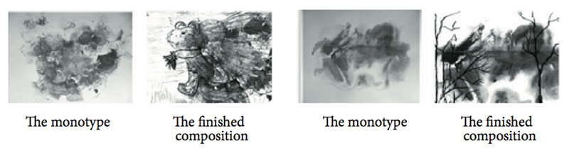
Figure 1. Examples of monotypes and finished compositions based on these artistic images
The EEG signals were recorded while the subjects were in a resting state with their eyes open, and during the four stages of creating an artistic image (viewing the monotypes, frustration, image creation, and thinking over the details). There were 21 scalp electrodes (Fpz, Fz, Cz, Pz, Oz, Fp1, Fp2, F3, F4, F7, F8, C3, C4, T3, T4, T5, T6, P3, P4, O1, O2), set according to a monopolar scheme with ipsilateral ear referents according to the International 10-20 System. Since the main part of the research was carried out when the participants’ eyes were open, a resting state with eyes open was used as the default state.
The stages of creating an artistic image were chosen by us to correspond to the stages of the creative process. The stage “Viewing of monotypes” corresponds to Preparation stage (definition of a problem, gathering of information, first attempts at solving the problem). At the Frustration stage, a person experiences negative emotions due to his/her inability to find a solution. The stage of “Image creation” corresponds to Insight (the sudden finding of a solution). And the stage of “Thinking over the details” corresponds to the stage of Verification (the creative idea is subjected to evaluation).
Only artifactless EEG samples, 10 seconds each, were analyzed. We analyzed EEG coherence for each functional trial at the following frequency bands: theta (4.00–8.00 Hz), alpha1 (8.00–10.5 Hz), alpha2 (10.5–13.00 Hz), and beta (13.00–35.00 Hz).
Coherent connections between pairs of electrodes for each frequency band were divided into the following groups: short-distance intra-hemispheric (4), long-distance intra-hemispheric (2), interhemispheric in the anterior and posterior cortex regions (2), cross-hemispheric (diagonal) (2), and interhemispheric symmetrical (between symmetrical pairs of leads) (2). Table 1 presents twelve kinds of analyzed coherent connections.
Average values were calculated for each kind of connection from each frequency band. The coherent connection was considered strong if its value was 0.7 or higher.
Statistical data processing
For statistical analysis, we used descriptive statistics and 3-way MANOVA (Multivariate analysis of variance). A comparative post hoc analysis according to the Fisher criteria was used as well. The processing was conducted with the help of STATISTICA 12.0 computer software.
Table 1. The kinds of coherent connections analyzed.

Results
Results of the descriptive analysis
A descriptive analysis of the average values of the coherence coefficients was carried out to determine the degree of cohesion of neural ensembles of various cerebral cortex regions in the representatives of the different professional groups at the different stages of the creative process. The results of the descriptive analysis for each frequency band are shown in Figures 2-5.
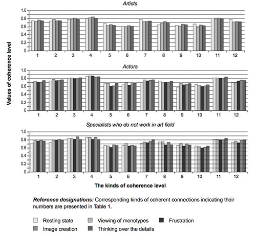
Figure 2. Average values of the analyzed coherent connections in the cortex of the research participants at different stages of artistic image creation at theta band
So, at the stage of viewing the monotypes, strong coherent connections are expressed in artists’ right anterior cortex regions, between the left anterior and posterior cortex regions, between the anterior left and right cortex regions, and between symmetrical leads of both hemispheres at theta band; between the posterior left and right cortex regions at theta and alpha1 bands; in the left anterior cortex regions at alpha2 band; in the right anterior cortex regions as well between the right anterior and posterior cortex regions at beta band (Fig. 2-5).
At the stage of image creation, high values of coherence were found in the artists’ anterior cortex regions of both hemispheres, and in the posterior and anterior cortex regions between brain hemispheres at theta band; in the posterior and ante rior cortex regions between hemispheres and between long-distance symmetrical leads of both hemispheres at alpha1 band; in the left anterior cortex regions at alpha2 band, and in the anterior cortex regions of both hemispheres at beta band.
Figure 3. Average values of the analyzed coherent connections in the cortex of the research participants at different stages of artistic image creation at alpha1 band
At the stage of thinking over the details, high values of coherence were found in the artists’ anterior cortex regions of both hemispheres, as well in the anterior and posterior cortex regions between brain hemispheres at theta band; in the posterior cortex regions between hemispheres at alpha1 band; in the left anterior cortex regions at alpha2 and beta bands (Fig. 2-5).
Strong coherent connections are expressed in actors at the stage of the viewing of monotypes in their left anterior cortex regions, between the right anterior and posterior cortex regions, and in the posterior cortex regions between brain hemispheres at theta band; in the left anterior cortex regions at alpha1 and alpha2 bands; between the anterior and posterior cortex regions in both hemispheres, and in the posterior cortex regions between hemispheres at alpha2 band; in the left anterior cortex regions as well as between the anterior and posterior cortex regions in both hemispheres at beta band (Fig. 2-5).
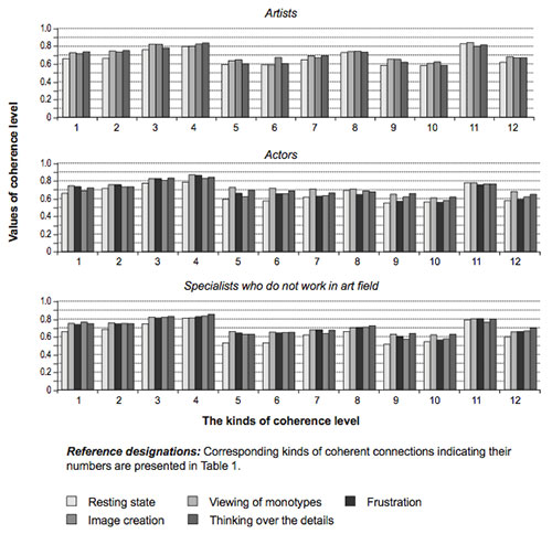
Figure 4. Average values of the analyzed coherent connections in the cortex of the research participants at different stages of artistic image creation at alpha2 band
At the stage of frustration, strong coherent connections are found in actors in the left anterior cortex regions at alpha1 band.
At the stage of image creation, the most expressive coherent connections in actors are located between the left and right posterior cortex regions at theta and alpha1 bands; in the left anterior cortex regions, and between the right anterior and posterior cortex regions at beta band.
At the stage of thinking over the details, high coherence values were found in actors between the anterior and posterior cortex regions of both hemispheres at theta band; between the posterior cortex regions of both hemispheres at alpha1 band; in the left anterior cortex regions at alpha2 band (Fig. 2-5).
The distribution of coherent connections in specialists who do not work in an artistic field, is the same as in artists and actors; only the power of the coherent links they have is higher.
So, the intra-hemispheric connections in the left and right posterior cortex regions, and interhemispheric symmetrical connections, especially in the anterior and posterior cortex regions, are dominant in these participants at all frequency bands.
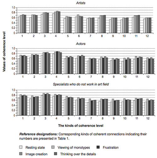
Figure 5. Average values of the analyzed coherent connections in the cortex of the research participants at different stages of artistic image creation at beta band
MANOVA results
We analyzed the frequency-specific effects of three factors: “GROUP”–1) artists, 2) actors, and 3) specialists who do not work in an artistic field; “TRIAL”–1) resting, 2) viewing of monotypes, 3) frustration, 4) image creation, and 5) thinking over the details; and “KIND OF COHERENT CONNECTION” (12).
As a result of the 3-way MANOVA, a number of frequency-specific effects of the factors GROUP, TRIAL, and KIND OF COHERENT CONNECTION, and their interactions in functional changes of EEG coherence, were revealed. The main effects of these factors’ interaction were significant in changes of functional EEG coherence for each frequency band, but with different degrees of significance (F (4, 193) = 5.89, p < .05 for theta band; F (1,851) = 9.64, p < .001 for alpha1 band; F (1,488) = 8.4, p < .001 for alpha2 band; and F (3, 785) = 6.93, p < .01 for beta band). The effects of the interaction of GROUP × TRIAL × KIND OF COHERENT CONNECTION were expressed to the greatest extent because their influence leads to significant changes in EEG coherence in the greatest number of different areas of the brain (p < .05). So we have carried out a comparative post hoc analysis (according to the Fisher criteria, p < .05) of values of EEG coherence in artists, actors and specialists not engaged in artistic sphere during different stages of artistic image creation.
The results of comparative post hoc analysis of values of EEG coherence in research participants during artistic image creation
As a result of comparative post hoc analysis, we revealed significant differences in the average values of coherence at the various stages of artistic image creation in the cortex of artists, actors and specialists who do not work in an artistic field (Tables 2–4).
Table 2. Average values of the coherent connections that are significantly changed at various stages of artistic image creation in the cortex of artists
|
Frequency bands |
Kinds of coherent connections |
Stages of an artistic image creation |
Overage values of coherent connections |
Significance levels |
|
|
Theta |
7 |
Resting state |
0.82692±0.15 |
p < .05 |
|
|
Viewing of monotypes |
0.73985±0.11 |
||||
|
12 |
Resting state |
0.82290±0.14 |
p < .05 |
||
|
Viewing of monotypes |
0.72993±0.12 |
||||
|
7 |
Resting state |
0.82692±0.15 |
p < .05 |
||
|
Image creation |
0.74310±0.11 |
||||
|
8 |
Resting state |
0.68041±0.16 |
p < .05 |
||
|
Image creation |
0.74066±0.1 |
||||
|
7 |
Resting state |
0.82692±0.15 |
p < .05 |
||
|
Thinking over the details |
0.75111±0.16 |
||||
|
Alpha2 |
11 |
Resting state |
0.83881±0.1 |
p < .05 |
|
|
Image creation |
0.78444±0.11 |
||||
|
Beta |
2 |
Resting state |
0.63535±0.12 |
p < .01 |
|
|
Viewing of monotypes |
0.71628±0.09 |
||||
|
4 |
Resting state |
0.77892±0.12 |
p < .05 |
||
|
Viewing of monotypes |
0.83244±0.09 |
||||
|
6 |
Resting state |
0.57514±0.07 |
p < .0001 |
||
|
Viewing of monotypes |
0.69066±0.09 |
||||
|
1 |
Resting state |
0.65876±0.12 |
p < .05 |
||
|
Image creation |
0.72086±0.09 |
||||
|
4 |
Resting state |
0.77892±0.12 |
p < .01 |
||
|
Image creation |
0.85520±0.09 |
||||
Note. Corresponding kinds of coherent connections indicating their numbers are presented in Table 1.
Table 3. Average values of the coherent connections that are significantly changed at various stages of artistic image creation in the cortex of actors
|
Frequency bands |
Kinds of coherent connections |
Stages of an artistic image creation |
Overage values of coherent connections |
Significance levels |
||
|
Alpha1 |
8 |
Resting state |
0.78712±0.07 |
p < .01 |
||
|
Frustration |
0.68833±0.08 |
|||||
|
Alpha2 |
4 |
Resting state |
0.60139±0.12 |
p < .01 |
||
|
Viewing of monotypes |
0.71054±0.14 |
|||||
|
3 |
Resting state |
0.62242±0.14 |
p < .01 |
|||
|
Viewing of monotypes |
0.71600±0.1 |
|||||
|
6 |
Resting state |
0.66756±0.13 |
p < .05 |
|||
|
Frustration |
0.74703±0.07 |
|||||
|
8 |
Resting state |
0.72367±0.08 |
p < .05 |
|||
|
Frustration |
0.64081±0.08 |
|||||
|
5 |
Viewing of monotypes |
0.74032±0.13 |
p < .01 |
|||
|
Frustration |
0.64446±0.14 |
|||||
|
4 |
Resting state |
0.60139±0.12 |
p < .05 |
|||
|
Thinking over the details |
0.67783±0.09 |
|||||
|
3 |
Viewing of monotypes |
0.71600±0.1 |
p < .01 |
|||
|
Image creation |
0.61692±0.1 |
|||||
|
5 |
Frustration |
0.64446±0.14 |
p < .05 |
|||
|
Thinking over the details |
0.71722±0.1 |
|||||
|
Beta |
2 |
Resting state |
0.67219±0.12 |
p < .001 |
||
|
Viewing of monotypes |
0.76660±0.1 |
|||||
|
1 |
Resting state |
0.63708±0.1 |
p < .001 |
|||
|
Viewing of monotypes |
0.73779±0.1 |
|||||
|
4 |
Resting state |
0.80950±0.11 |
p < .05 |
|||
|
Viewing of monotypes |
0.86891±0.08 |
|||||
|
3 |
Resting state |
0.76855±0.13 |
p < .01 |
|||
|
Viewing of monotypes |
0.84683±0.09 |
|||||
|
6 |
Resting state |
0.63236±0.11 |
p < .01 |
|||
|
Viewing of monotypes |
0.72063±0.1 |
|||||
|
5 |
Resting state |
0.62956±0.09 |
p < .05 |
|||
|
Viewing of monotypes |
0.68766±0.13 |
|||||
|
2 |
Resting state |
0.67219±0.12 |
p < .05 |
|||
|
Frustration |
0.74095±0.09 |
|||||
|
1 |
Resting state |
0.63708±0.1 |
p < .01 |
|||
|
Frustration |
0.71753±0.08 |
|||||
|
4 |
Resting state |
0.80950±0.11 |
p < .05 |
|||
|
Frustration |
0.87904±0.08 |
|||||
|
3 |
Resting state |
0.76855±0.13 |
p < .01 |
|||
|
Frustration |
0.84491±0.11 |
|||||
|
6 |
Resting state |
0.63236±0.11 |
p < .05 |
|||
|
Frustration |
0.69787±0.07 |
|||||
|
2 |
Resting state |
0.67219±0.12 |
p < .001 |
|||
|
Image creation |
0.77254±0.08 |
|||||
|
1 |
Resting state |
0.63708±0.1 |
p < .001 |
|||
|
Image creation |
0.73326±0.1 |
|||||
|
4 |
Resting state |
0.80950±0.11 |
p < .05 |
|||
|
Image creation |
0.87290±0.09 |
|||||
|
3 |
Resting state |
0.76855±0.13 |
p < .01 |
|||
|
Image creation |
0.84887±0.1 |
|||||
|
6 |
Resting state |
0.63236±0.11 |
p < .001 |
|||
|
Image creation |
0.74004±0.07 |
|||||
|
5 |
Resting state |
0.62956±0.09 |
p < .001 |
|||
|
Image creation |
0.72607±0.11 |
|||||
|
6 |
Resting state |
0.63236±0.11 |
p < .05 |
|||
|
Thinking over the details |
0.69905±0.09 |
|||||
Note. Corresponding kinds of coherent connections indicating their numbers are presented in Table 1.
Table 4. Average values of the coherent connections that are significantly changed at the stages of artistic image creation in the cortex of specialists who do not work in an artistic field
|
Frequency bands |
Kinds of coherent connections |
Stages of an artistic image creation |
Overage values of coherent connections |
Significance levels |
|
Theta |
8 |
Resting state |
0.77829±0.19 |
p < .05 |
|
Frustration |
0.68147±0.11 |
|||
|
9 |
Resting state |
0.73860±0.15 |
p < .01 |
|
|
Frustration |
0.65353±0.15 |
|||
|
9 |
Viewing of monotypes |
0.72056±0.13 |
p < .05 |
|
|
Frustration |
0.65353±0.15 |
|||
|
8 |
Resting state |
0.77829±0.19 |
p < .05 |
|
|
Thinking over the details |
0.67725±0.14 |
|||
|
Alpha2 |
1 |
Resting state |
0.67920±0.15 |
p < .05 |
|
Image creation |
0.75586±0.12 |
|||
|
Beta |
2 |
Resting state |
0.69006±0.08 |
p < .01 |
|
Viewing of monotypes |
0.77404±0.1 |
|||
|
1 |
Resting state |
0.71441±0.1 |
p < .001 |
|
|
Viewing of monotypes |
0.82137±0.09 |
|||
|
6 |
Resting state |
0.64222±0.06 |
p < .05 |
|
|
Viewing of monotypes |
0.71355±0.09 |
|||
|
5 |
Resting state |
0.65368±0.11 |
p < .001 |
|
|
Viewing of monotypes |
0.76124±0.1 |
|||
|
1 |
Resting state |
0.71441±0.09 |
p < .05 |
|
|
Frustration |
0.77976±0.1 |
|||
|
1 |
Resting state |
0.71441±0.09 |
p < .01 |
|
|
Image creation |
0.80359±0.1 |
|||
|
1 |
Resting state |
0.71441±0.09 |
p < .05 |
|
|
Thinking over the details |
0.77702±0.11 |
Note. Corresponding kinds of coherent connections indicating their numbers are presented in Table 1.
The high values of intra- and interhemispheric coherence between certain brain regions were revealed in all research participants at theta band. In addition, the dynamics of their distribution varied at different stages of artistic image creation (Fig. 6).
A weakening of short-distance coherent links within the left hemisphere at theta band was observed at the stage of viewing of monotypes, compared to a resting state in representatives of artistic professions (p < .05). A decrease in interhemispheric connections was also revealed: artists had less interhemispheric long-distance symmetrical connections; actors had less interhemispheric anterior, posterior and long-distance symmetrical connections (p < .05). In representatives of both creative groups, there were distinct short-distance connections in the right hemisphere at the stage of viewing of monotypes; artists also demonstrated interhemispheric anterior connections (p < .05) (Tables 2, 3).
Artists exhibit a strengthening of short-distance connections in the left hemisphere (p < .05) and posterior interhemispheric connections (p < .01) at theta band at the stage of image creation and thinking over the details of composition. The stage of image creation is characterized by an increased intensity of short-distance left hemisphere connections, and interhemispheric anterior and long-distance symmetrical connections in the actors’ brains. Increased intensity of the coherence of interhemispheric posterior and short-distance symmetrical connections (p < .05) is typical for the stage of thinking over the details of composition (Tables 2, 3).
The EEG did not reveal any changes in the cortex’s functional state in specialists who do not work in an artistic field, at theta band during the image creation process compared to the resting state; high values of intra- and interhemispheric coherence were observed during the whole course of the research study (Table 4).
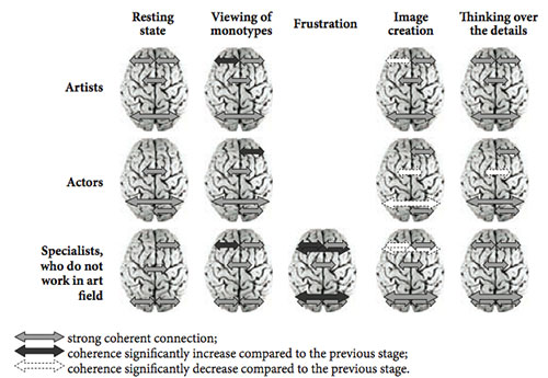
Figure 6. Significant differences in the strength of functional cortex connections at theta band in representatives of different professional groups at different stages of artistic image creation (p < .05)
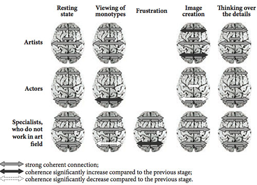
Figure 7. Significant differences in the strength of functional cortex connections at alpha1 band in representatives of different professional groups at different stages of artistic image creation (p < .05)
A high level of intra-hemispheric interaction in both hemispheres was identified at all stages of artistic image creation in all research participants at alpha1 band (Fig.7). Meanwhile, different stages of the creative process of representatives of each professional group are characterized by enhanced interhemispheric interaction in the posterior brain cortex at alpha1 band (p < .01): The artists have it at the stage of image creation, actors at the stage of viewing of monotypes; the specialists who do not work in an artistic field, demonstrate it at the stage of frustration (Tables 2-4). Increased intensity of coherence of the anterior interhemispheric connections is also observed in artists’ brains at the stage of image creation (p < .01).
Analysis of the coherence at alpha2 band showed values of intra- and interhemispheric connections between certain brain cortex regions during artistic image creation that varied for the representatives of each professional group under study (Fig. 8). artists have high values of coherence in short-distance left hemispheric connections when they are viewing monotypes; however, the significance of these values decreases at the stage of image creation (p < .05).
Low values of coherence in the anterior connections of the left hemisphere (p < .05) and high values in short-distance connections of the right hemisphere (p < .01) are inherent in the actors’ brain throughout the creative process (Table 3).
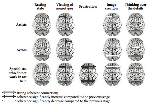
Figure 8. Significant differences in the strength of functional cortex connections at alpha2 band in representatives of different professional groups at different stages of artistic image creation (p < .05)
The specialists who do not work in an artistic field, have increased short-distance anterior connections in the left hemisphere at the stage of viewing of mono-types, compared with the resting state, and increased intensity of interhemispheric anterior and posterior connections at the stage of frustration (p < .05). The stages of image creation and thinking over the details are characterized by a decreased intensity of short-distance anterior left- and interhemispheric connections (p < .05) (Table 4).
At beta band, the dynamics of the distribution of coherence connections is similar among subjects in all groups. (Fig. 9). At all stages of artistic image creation, a high level of intra-hemispheric interaction in both hemispheres (p < .01) and a low level of interhemispheric interaction (p < .01) were revealed. This result can serve as a proof of independent and parallel functioning of brain hemispheres, and separate processing of graphic information (Behtereva & Nagornova, 2007; Dikaya et al., 2016).
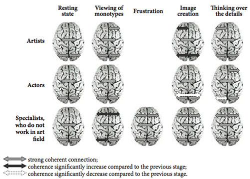
Figure 9. Significant differences in the strength of the functional cortex connections at beta band in representatives of different professional groups at different stages of artistic image creation (p < .05)
As a result of the research, neurophysiological correlates of artistic image creation were studied for representatives of different artistic professions (artists, actors) at different stages of creative process.
Discussion
Contemporary researchers connect the functional role of theta rhythm not only with the regulation of emotions, but consider that it can also be a sign of directing attention outward; the readiness of a test subject to perform a task; intensity of effort; and the creation of conditions of increased neuron plasticity (Korobejnikova, 2011). On the basis of the above, we concluded that the indicators of coherence in theta band of the research participants’ EEG can reflect the intensity of effort exerted to create an artistic image, and can be a sign of concentration on directing attention outward.
Hence, the local zones of intensity of effort were revealed at theta band at the stage of viewing of monotypes—in the right posterior area that is engaged in processing image information, as well as in integrating images into one coherent spatial image. The left-hemispheric functional connections—at the stages of image creation and thinking over its details—reflect the engagement of information analysis mechanisms. The detected enhancement of coherent connections in the right frontal area, and the interhemispheric anterior connections at different stages of artistic image creation at theta band, can represent peculiarities of the engagement of active attention mechanisms in different artistic professionals.
Scientists declare that the functional role of alpha1 rhythm is connected with a general state of activation, the actualization of intuitive processes, internal information processing, and inhibition of information that is irrelevant to fulfilling the current task (Behtereva & Nagornova, 2007; Farber, Machinskaja, Kurganskij, & Petrenko, 2014; Fink & Benedek, 2014; Fink, Schwab, & Papousek, 2011).
The enhancement of posterior interhemispheric coherent connections that was revealed by our research coheres with contemporary views on changes in synchronization of alpha rhythm in modally-specific cortex zones (Farber et al., 2014), and with the opinion that an enhancement of alpha rhythm reflects inhibition of any distracting and interfering information flowing from the visual system (Fink et al., 2011). The increased intensity of frontal interhemispheric connections in artists’ brains that was detected in the course of the research at the stage of image creation, can also indicate suppression of cognitive processes that are not directly related to the current tasks.
Contemporary scientists assert that the functional role of alpha2 rhythm relates to the specific character of information processing while solving cognitive tasks. It is believed that the right frontal region is involved in spontaneous production of non-verbal representations, and that the left one carries out control, gives additional assessment and analysis, and provides for the purposeful extraction of information from episodic and semantic memory (Razumnikova, Finikov, 2011). On the basis of the above, we concluded that the process of spontaneous image creation is launched later in the brains of the artists; while mechanisms of analysis and assessment of the given material (monotypes) dominate during the viewing of monotypes. The total creative process of actors is based on the search for possible associations, and spontaneous image generation.
Thus, representatives of all the artistic professions exhibited:
- strong short-distance intra-hemispheric connections in theta band, the presence of which can reflect the existence of local zones of intensity of effort: at the stage of viewing of monotypes, in the right posterior region; at the stage of image generation and thinking over its details, in the left hemisphere;
- strong interhemispheric connections at alpha1 band, which can reflect the suppression of cognitive processes that are not directly related to the current task: actors have it at the stage of viewing of monotypes; artists, at the stage of image creation;
- strong intra-hemispheric connections at beta band at all stages of artistic image creation and a decrease of interaction between hemispheres while searching for remote imaginative associations—i.e. conceiving the picture concept.
The actors demonstrate strong interhemispheric frontal connections in theta band, which likely provide the peculiarities of the involvement of voluntary attention mechanisms. The distribution of functional connections of the brain cortex of the actors at alpha2 band has mainly right-hemispheric localization. Moreover, it provides a simultaneous method of information processing for efficiently generating new images. The artists have equally distinct interhemispheric frontal connections at all stages of artistic image creation at theta-band (peculiarities of the involvement of voluntary attention mechanisms); increased coherence in posterior interhemispheric connections are observed at the stages of image creation and thinking over its details.
The distribution of functional cortex connections in alpha2 band is connected with equal involvement of right and left hemispheres during the process of imaginative creative activity in artists’ brains; spontaneous generation of images and the mental design of artistic composition are promoted on this basis.
Conclusion
The results of this research permit us to draw the following conclusions:
- The character of the brain’s functioning during imaginative creative activity proves that all the females aged 23-27 participating in the research were highly emotionally involved in the creative process, and demonstrated similar levels of brain cortex activity at all the stages of the process.
- The differences in brain cortex functioning when an artistic image is created by artists (close interaction of brain hemispheres) or actors (dominance of right hemisphere activity) reflect different strategies for carrying out imaginative creative work by representatives of these professions.
- The analysis of the supplied monotypes and their assessment precedes the spontaneous and insightful creation of images by the artists. They create the general idea of an artistic image in their mind’s eye, and then they think over the possibility of combining this idea and the supplied monotype into a holistic image.
- A distinct, simultaneous way of processing the information provided for the efficient generation of new images is typical for actors. Their creative process is based on the search for possible associations, and spontaneous image generation.
These results can be used in education to develop creative and artistic potential in students, and in vocational guidance and training for artists, actors and other representatives of the artistic sphere. Our results may also be used in clinical psychology and medicine for psychotherapy and art therapy, for personal psycho-correction (to release the patients’ creative potential), and for treating mental and somatic diseases as well.
Perspectives for further investigation of the problem include studying the main neurophysiological correlates of the creative process per se independent of a specific professional domain. It is not surprising for an artist to show different brain activity patterns from those of a non-artist, when he/she is thinking about an artistic image. The question remains whether an artist shows the same brain activity when thinking about an artistic image as the actor does when thinking about the image of an actor’s role, or as a musician does when thinking about music, for example. The results obtained demonstrate the need for current models of the neural basis of the creative process to be developed further.
Acknowledgements
This research was supported by the Ministry of Education and Science of the Russian Federation, project № 2141.
References
Annett, M. (1970). A classification of hand preference by association analysis. British Journal of Psychology, 61(3), 303–321. doi: 10.1111/j.2044-8295.1970.tb01248.x
Asari, T., Konishi, S., Jimura, K., Chikazoe, J., Nakamura, N., & Miyashita, Y. (2008). Right temporopolar activation associated with unique perception. Neuroimage, 41(1), 145–152. doi: 10.1016/j.neuroimage.2008.01.059
Bechtereva, N. P., & Nagornova, Z. V. (2007). Changes in EEG coherence during tests for nonverbal (figurative) creativity. Human Physiology, 33(5), 515-523. doi: 10.1134/ S0362119707050015
Bhattacharya, J., & Petsche, H. (2005). Drawing on mind’s canvas: Differences in cortical integration patterns between artists and non-artists. Human Brain Mapping, 26(1), 1–14. doi: 10.1002/hbm.20104
Bondareva, O. V. (2009). Specifika prepodavanija i osvoenija discipliny “Hudozhestvennaja grafika i graficheskaja kompozicija”» studentam-dizajneram na hudozhestvenno-graficheskih fakul’tetah [The specific character of teaching “Art graphics and graphic composition” to students with design major at arts and graphic departments and its development].
Materialy Mezhdunarodnoj nauchno-prakticheskoj konferencii: formirovanie professional’nyh kompetencij v vysshem obrazovanii v XXI veke [Proceedings of International Scientific-Practical Conference: Development of Professional Competencies in Higher Education in the 21st century]. (pp. 155–159). Moscow – Elecrostal: New Humanities Institute.
Dietrich, A., & Kanso, R. (2010). A review of EEG, ERP, and neuroimaging studies of creativity and insight. Psychological Bulletin, 136(5), 822–848. doi: 10.1037/a0019749
Dikaya, L. A., & Dikiy, I. S. (2015). Tvorcheskij mozg [Creative brain]. Rostov-on-Don: Southern Federal University Press.
Dikaya, L. A., Dikiy, I. S., Karpova, V. V., & Lavreshina, A. Y. (2016). Artists’ Brain Cortex Functional Organization at the Insight Stage of Creative Problem Solving. International Journal of Psychophysiology, 108, 132–133. doi: 10.1016/j.ijpsycho.2016.07.390
Dikaya, L. A., Naumova, M. I., & Naumov, I.V. (2015). Psihofisiologicheskie korreliaty myslennogo ispolneniya improvizirovannogo tantsa [Psychophysiological correlates of mental performance of the improvisation dance]. Rossijskij Psihologicheskij Zhurnal [Russian Psychological Journal], 12(4), 110–126.
Dikaya, L. A., & Skirtach, I. A. (2015). Neurophysiological correlates of musical creativity: The example of improvisation. Psychology in Russia, 8(3), 84–98. doi: 10.11621/pir.2015.0307
Dikiy, I. S., Dikaya, L. A., & Karpova, V. V. (2016). Brain Correlates of the Artistic Image Creation by Artists and Actors. International Journal of Psychophysiology, 108, 131. doi: 10.1016/j. ijpsycho.2016.07.386
Dikiy, I. S., Dikaya, L. A., & Skirtach, I. A. (2014). Interhemispheric functional organization of brain cortex in musicians during improvisation. International Journal of Psychophysiology, 94(2), 127. doi: 10.1016/j.ijpsycho.2014.08.606
Farber, D. A., Machinskaja, R. I., Kurganskij, A. V., & Petrenko, N. E. (2014). Funkcional’naja organizacija mozga v period podgotovki k opoznaniju fragmentarnyh izobrazhenij [Functional organization of the brain while preparing for the identification of fragmented images] Zhurnal Vysshey Nervnoy Deyatelnosti [Journal of Higher Nervous Activity], 64(2), 190–200. doi: 10.7868/S0044467714020075
Fink, A., & Benedek, M. (2014). EEG alpha power and creative ideation. Neuroscience & Biobehavioral Reviews, 44, 111–123. doi: 10.1016/j.neubiorev.2012.12.002
Fink, A., Graif, B., & Neubauer, A. C. (2009). Brain correlates underlying creative thinking: EEG alpha activity in professional vs. novice dancers. NeuroImage, 46(3), 854–862. doi: 10.1016/j. neuroimage.2009.02.036
Fink, A., Schwab, D., & Papousek, I. (2011). Sensitivity of EEG upper alpha activity to cognitive and affective creativity interventions. International Journal of Psychophysiology, 82(3), 233–239. doi:10.1016/j.ijpsycho.2011.09.003
Gibson, C., Folley, B. S., & Park, S. (2009). Enhanced divergent thinking and creativity in musicians: A behavioral and near-infrared spectroscopy study. Brain and Cognition, 69(1), 162-169. doi: 10.1016/j.bandc.2008.07.009
Gonen-Yaacovi, G., de Souza, L. C., Levy, R., Urbanski, M., Josse, G., & Volle, E. (2013). Rostral and caudal prefrontal contribution to creativity: A meta-analysis of functional imaging data. Frontiers in Human Neuroscience, 7, 465. doi: 10.3389/fnhum.2013.00465
Jaušovec, N. (2000). Differences in cognitive processes between gifted, intelligent, creative, and average individuals while solving complex problems: an EEG study. Intelligence, 28(3), 213237. doi:10.1016/S0160-2896(00)00037-4.
Jin, S. H., Kwon, Y. J., Jeong, J. S., Kwon, S. W., & Shin, D. H. (2006). Differences in brain information transmission between gifted and normal children during scientific hypothesis generation. Brain and Cognition, 62(3), 191–197. doi: 10.1016/j.bandc.2006.05.001
Jung, R. E., Mead, B. S., Carrasco, J., & Flores, R. A. (2013). The structure of creative cognition in the human brain. Frontiers in Human Neuroscience, 7, 330. doi: 10.3389/fnhum.2013.00330
Jung-Beeman, M., Bowden, E. M., Haberman, J., Frymiare, J. L., Arambel-Liu, S., Greenblatt, R., & Kounios, J. (2004). Neural activity when people solve verbal problems with insight. PLoS Biol, 2(4), e97. doi:10.1371/journal.pbio.0020097
Korobeynikova, I. I. (2011). Svyaz prostranstvennoy sinkhronizatsii teta-diapazona EEG cheloveka s raznoy uspeshnost’yu vypolneniya zritelno-prostanstvennykh zadach [The relationship of the spatial synchronization of theta-band EEG with the successful implementation of various spatio-visual tasks]. Fiziologiya Cheloveka [Human Physiology], 37(5), 26–34. doi: 10.7868/S004446771306004X
Kounios, J., & Beeman, M. (2014). The cognitive neuroscience of insight. Psychology, 65(1), 71. doi: 10.1146/annurev-psych-010213-115154
Kowatari, Y., Lee, S. H., Yamamura, H., Nagamori, Y., Levy, P., Yamane, S., & Yamamoto, M. (2009). Neural networks involved in artistic creativity. Human Brain Mapping, 30(5), 1678– 1690. doi: 10.1002/hbm.20633
Limb, C. J., & Braun, A. R. (2008). Neural substrates of spontaneous musical performance: An fMRI study of jazz improvisation. PLoS ONE, 3(2), e1679. doi: 10.1371/journal. pone.0001679
Liu, S., Chow, H. M., Xu, Y., Erkkinen, M. G., Swett, K. E., Eagle, M. W., & Braun, A. R. (2012). Neural correlates of lyrical improvisation: An fMRI study of freestyle rap. Scientific Reports, 2, 834. doi: 10.1038/srep00834
Mayseless, N., & Shamay-Tsoory, S. G. (2015). Enhancing verbal creativity: Modulating creativity by altering the balance between right and left inferior frontal gyrus with tDCS. Neuroscience, 291, 167-176. doi: 10.1016/j.neuroscience.2015.01.061
Pereda, E., Rahman, S., & Bhattacharya, J. (2014). The structure of functional brain EEG network is sensitive to genre-specific musical creativity: A study on jazz and classical piano music performance. International Journal of Psychophysiology, 2(94), 161. doi:10.1016/j. ijpsycho.2014.08.705
Pinho, A. L., de Manzano, Ö., Fransson, P., Eriksson, H., & Ullén, F. (2014). Connecting to create: Expertise in musical improvisation is associated with increased functional connectivity between premotor and prefrontal areas. The Journal of Neuroscience, 34(18), 6156-6163. doi: 10.1523/JNEUROSCI.4769-13.2014
Razumnikova, O. M., & Finikov, S. B. (2011). Otrazhenie social’noj kreativnosti v osobennostjah aktivacii kory na chastotah del’ta-, al’fa2- i gamma2- ritmov. [Reflection of the social creativity in the activation features of the cortex at frequencies of the delta, and alfa2- gamma2- rhythms]. Zhurnal vysshej nervnoj dejatel’nosti [Journal of Higher Nervous Activity], 61 (6), 706-715.
Razumnikova, O. M., & Yashanina, A. A. (2014). Personality specific differences in EEG reactivity on convergent and divergent thinking. International Journal of Psychophysiology, 94(2), 160. doi: 10.1016/j.ijpsycho.2014.08.702
Rodionov, A. R. (2013). Mozgovye mehanizmy voobrazhenija pri vypolnenii verbal’nyh tvorcheskih zadach [Brain mechanisms of imagination in the process of carrying out verbal creative tasks]. Fiziologija Cheloveka [Human Physiology], 39(3), 35-45. doi:10.7868/ S0131164613030168.
Starchenko, M. G., Kireev, M. V., & Medvedev, S. V. (2014). Brain organization in creative thinking. International Journal of Psychophysiology, 94(2), 160. doi: 10.1016/j. ijpsycho.2014.08.703
Villarreal, M. F., Cerquetti, D., Caruso, S., Aranguren, V. S. L., Gerschcovich, E. R., Frega, A. L., & Leiguarda, R. C. (2013). Neural correlates of musical creativity: differences between high and low creative subjects. PloS ONE, 8(9), e75427. doi: 10.1371/journal.pone.0075427
Vygotsky, L. S. (2005). Problema razvitiya I raspada vysshikh psikhicheskikh funktsiy [The issue of development and deterioration of higher mental functions]. In Psihologiya razvitiya cheloveka [Psychology of human development]. Moscow: Smysl. (Original work published 1934)
Wiggins, G. A., & Bhattacharya, J. (2014). Mind the gap: an attempt to bridge computational and neuroscientific approaches to study creativity. Frontiers in Human Neuroscience, 8. doi: 10.3389/fnhum.2014.00540
Zaidel, D. W. (2014). Creativity, brain, and art: Biological and neurological considerations. Frontiers in Human Neuroscience, 8, 389. doi: 10.3389/fnhum.2014.00389To cite this article: Dikaya L. A., Dikiy I. S., Karpova V. V., Lavreshina A. Yu. (2016). Neurophysiological correlates of artistic image creation by representatives of artistic professions. Psychology in Russia: State of the Art, 9(4), 51-72.
The journal content is licensed with CC BY-NC “Attribution-NonCommercial” Creative Commons license.