Vector Encoding of Light Intensity in Neural Networks of Visual System
Abstract
Intracellular research of land snail Helix lucorum L. eye demonstrates two types of visual cells responding to flashes of white light by slow sustained depolarization (D-type) and by slow sustained hyperpolarization (H-type), respectively. Peaks of spectral sensitivity of both cell types at 465-500 nm coincide with peak of spectral sensitivity of photopigment "rhodopsin". The two-channel vector model of achromatic vision of snail is proposed. According to the model, responses of D- and H-cells constitute two-dimensional 'excitation vector' of constant length, the direction of which is the code of light intensity. The two-dimensional vector model of light encoding in snails' eye is analogous with achromatic vision models of achromatic vision in vertebrates based on psychophysical and neurophysiological data in fish, rabbit, monkey and human. So, intracellular data in snail taken together with data on vertebrate animals testify in favor of the hypothesis that 2-dimensioanl module of "bright" and "dark" cells is the universal opponent mechanism of "vector encoding" of light intensity in neuronal nets for vision.
Themes: Psychophysiology
Pages: 309-318
DOI: 10.11621/pir.2008.0020
Keywords: psychophysiology, methodology
I. Introduction
Psychophysical, behavioral and neurophysiological researches of light and color brightness perception in vertebrate animals show that there are two types of neurons, opponent to each other in differentiation of brightness gradations (Jung, 1973; DeValois, DeValois, 1975; Chernorizov, Sokolov, 2001; Sokolov, 2003; Izmailov et al., 2006; Chernorizov, 2006, 2007). Neurons of the first type gradually increase their activity while light intensity increases, and reduce it while light intensity reduces. These cells are called
"B-type cells". The neurons of the second type respond to changes of brightness in strictly opposite way. They are known as "D-rype cells". Taking into consideration high vertebrates, B- and D-neurons are found in LGN and in visual cortex of monkey, rabbit and cat (Fig. 1, 2). As for lower vertebrates (fish, amphibia) there are analogues to B- and D-neurons cells, which are known as cells of ON- and OFF-types (according to the neurons classification offered by Hartline, 1940). These cells are already found in retina at the level of bipolar cells and ganglion cells (Fig. 3). According to the___________________ proposed by prof. E. Sokolov and proved in psychophysical experiments with human, the characteristics of B- and D-neurons correspond topredetectors forming "2-dimensionalneuronal module for brightness coding" (Sokolov, 2000, 2003, 2006). Coding of brightness in such a module is carried out by 2-dimensional "vector of excitation". Components of this vector are responses' values of B- and D-neurons. One can consider such kind of an "excitation vector" as an "encoding vector of lightness" at the level of predetector neurons (Fig. 4). This 2-dimensional module incorporates as a subset into the color vision system in vertebrates (Chernorizov, Sokolov, 2001; Sokolov, 2003, 2006).
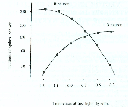
Figure 1. Opponent characteristics of B- and D-cells in cat's visual cortex (Jung, 1973).
It raises a reasonable question: what are the neural mechanisms of achromatic vision of invertebrates and how do they correspond with neural mechanisms of brightness in vertebrates? For answering this question, we have chosen mollusk//e//xpomatia L. (grape snail) as an object for research. Retina of a mollusk's eye contains only one photopigment - rhodopsin. For this reason snail can distinguish gradations only of light intensity but not of color. Thus, simply organized visual system of snail represents convenient object for studying neural mechanisms of brightness in invertebrates' animals.
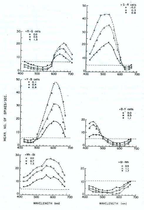
Figure 2. Spectral characteristics of B- and D-neurons in LGN of monkey designated as +Wh-Bl and +Bl-Wh cells in low pair of curves (DeValois, DeValois, 1975).
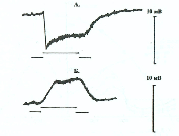
Figure 3. Responses of bipolar cells of OFF- (A) and ON-types (B) in carp retina to flashes of white light in the centre their receptive field (Chernorizov, Sokolov, 2001).
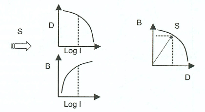
Figure 4. Hypothesis of vector encoding of light intensity in visual system by combination of excitations in neurons of B- and D-types (Chernorizov, Sokolov, 2001; Sokolov, 2003). According to the hypothesis of vector encoding, the characteristics of B- and D-neurons correspond to predetectors forming "2-dimentional neuronal module for brightness coding". Coding of brightness in such a module is carried out by 2-dimensional "vector of excitation". Components of this vector are responses' values of B- and D-neurons.
II. Experimental Data of Intracellular Research
In this report we analyze the experimental intracellular data concerning responses of photosensitive cells of snail's retina. These data testify to existence of "light" and "dark" cells that are analogous, accordingly, to Band D-neurons in visual system of vertebrates.
1. Experimental technique
Experiments were made on dark-adapted half-intact preparation "eye glass - optical nerve - cerebral ganglion". Access to retina was reached by opening of an eye ball and removal of a crystalline lens. For light stimulation we used (1) the diffusive flashes of white light and (2) equiquantum monochromatic radiations of different wavelengths (in 400-700 nanometers interval). Duration of stimulus was 1 second, intensity varied within the limits of 1,5 lg. units with maximum that equals 5,0 • 105 erg • snr2 • s"1 (0,0 lg units) for monochromatic radiations at a preparation level. The experimental setup for intracellular recording included Intracellular amplifier MEZ-8201 (Nihon Kohden), two-beam Oscilloscope VC-10 (Nihon Kohden) (0 + 1000 Hz) and Personal computer IBM with 16-channel analogue-digital converter (frequency of digitizing - 400 Hz). As microelectrodes we used glass ultrathin tubes with diameter of tip less than 1-2 micron and resistance of about 40-80 MOm. Collection and analysis of experimental data was implemented with the help of computer program "CONAN" (Kulaichev, 2002).
2. Experimental results. Types of photosensitive neurons in snail retina and their characteristics.
In 47 experiments on 38 animals' intracellular and, in some cases, extracellular recordings the responses of 53 cells were obtained and analyzed. In accordance with the sign of light response (sustained depolarization or sustained hyperpolarization) most of the registered cells have been divided into two groups. White light or any monochromatic color (D-type cells) depolarized the cells in the first group (Fig. 5). The cells in the second group responded to light of any wavelength or white light by hyperpolarization (H-type cells; Fig. 6). The functions of spectral responses of both H- and D-cells finely corresponded to spectral characteristic of rhodopsin (see also: Chernorizov et al., 1992). Amplitudes of responses linearly corresponded with the logarithm of light intensity (Fig. 7).
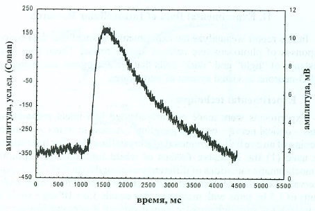
Figure 5. The response of D-cell to diffuse flash of white light. Along axis X - duration of light stimulation, ms; along axis Y - amplitude of response, mV (on the right), and artificial units of computer program (on the left). Duration of light flash is 1000 ms. Onset of light stimuli - left upward arrow, offset of light stimuli - right upward arrow.
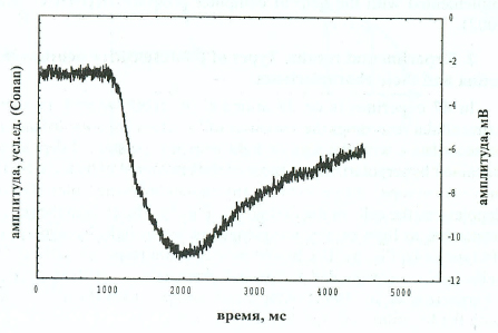
Figure 6. The response of H-cell to diffuse flash of white light. Designations as to Fig.5.
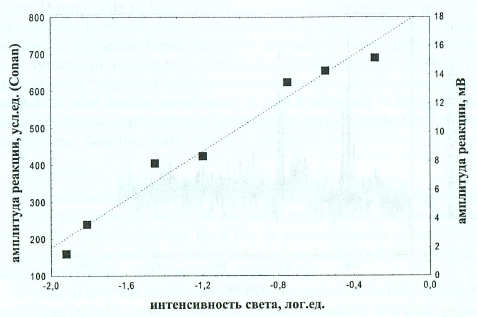
Figure 7. Amplitude of H-cell responses as a function of light intensity. The dark level of H-cell membrane potential equaled -5 mV. Designations as to Fig.5.
Besides D- and H-cells we have also found out 18 cells, which answered light flashes not with sustained responses but with spike discharges. Among them the following cells were there:
- Cells with equally well-expressed phasic impulse responses to onset and offset of diffuse flash light (ON/OFF-elements in vertebrate retinas in classification by Hartline, 1940) (Fig. 8).
- Cells with poorly expressed impulse discharge on light's turning on and with powerful discharge on light's turning off {OFF-elements, after Hartline, 1940).
- Cells with reverse correlation of discharge expressiveness on turning on and turning off the light (ON-elements, after Hartline, 1940).
III. Discussion
- Cells in situ and in isole. H- and D-type cells obtained in our research are completely corresponding with two analogous types of photosensitive cells of snail's retina discovered in experiments on the enzymatically isolated cells (Shekchter, Gretchenko, 2008).
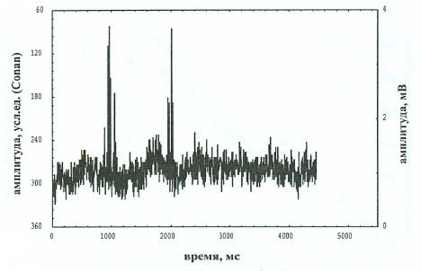
Figure 8. The response of ON/OFF-cell in mollusk retina to white light. Designations as to Fig. 5
- Morphological types of retinal cells generating FI- and D-responses: hypothesis about existence of two types of photoreceptors in a snail retina. There a question arises on the anatomy of "brightness cells" in snail retina: what are the morphological types of retinal cells generating the D- and H-responses? Whether they are photoreceptors, interneurons, pigment cells or glial cells? Considering all the data available now about features of invertebrates photoreception (for review see: Musio, 2001), we propose a hypothesis about presence of two photoreceptors types in a snail's eye retina: depolarized (rabdomeric type) and hyperpolarized (ciliary type) in response to light. Such combination of two morphologically and functionally different types of photoreceptors is unique: up to now it is only revealed in retinas of two mollusks - Lima scarba and Pecten irradians (Nasi, 1991 А, В, C; Gomez Nasi, 1998, 2000; Musio, 2001).
- The cells of H- and D-types in mollusk retina are the analogues of Band D-type neurons in vertebrates' visual system (?). Revealed in our research cells of H-type are hyperpolarized by light and depolarized in darkness. D-type cells have reverse type of responses. The responses of H- and D-cells in mollusk retina look like responses of ON- and OFF- bipolar cells in lower vertebrates (compare Fig. 3 and Fig. 5, 6). It gives us a basis for considering H- and D-type cells to be analogues to "dark" (D-type) and "bright" (B-type) neurons in visual system of vertebrates (Jung, 1973). Such analogy emphasizes a possible basic generality in mechanisms of light coding in visual systems of vertebrate and invertebrate animals. This "generality" is based on using "opponent" cells in mechanisms of coding brightness of light (mollusk: H- and D-cells; vertebrates: B- and D-neurons). "Opponent" cells generate 2-dimensional vector of excitation as early as at the level of retina (Chernorizov, Sokolov, 2001; Sokolov, 2003).
Conclusions
- Experiments with intracellular recording reveal two different types of cells in the retina of snail with opponent responses to diffuse flashes of light. Under light stimulation by white or monochromatic stimuli D-cells demonstrate sustained depolarization (that sometimes accomplished by spikes) and H-cells - sustained hyperpolarization.
- Peak of spectral sensitivity for both H- and D-cells falls at 470-500 nm area and coincides with the area of maximal sensitivity of a photopigment "rhodopsin".
- Dependence of amplitude of D- and H-cells responses on the logarithm of light intensity was found out to be well approximated by linear function and in such a way submits to Fehner's law.
- Intracellular data in snail taken together with data on vertebrate animals testify in favor of the hypothesis that 2-dimensioanl module of "bright" and "dark" cells is the universal opponent mechanism of "vector encoding" of light intensity in neuronal nets for vision.
References
Chernorizov, A.M. Vector encoding of light intensity in neuronal nets of snail eye // Intern. J. Psychophysiol. 2006. V. 61. N 3. P. 307-308.
Chernorizov, A.M., Shekhter, E.D., Arakelov, G.G., Zimachev, M.M. Vision in snail: spectral sensitivity of dark-adapted eye // Zh. VND. 1992. V 42. N 6. P. 1150-1156. (in Russian).
Chernorizov, A.M., Sokolov, E.N. Vector encoding of color in bipolar cells of carp retina // Vestnik MGU. Seria 14. Psyhologiya. 2001. N 1. P. 1233. (in Russian).
Chernorizov, A.M., Zimachev, M.M., Shekhter, E.D., Garusev A.V. Mechanisms of Achromatic Vision in Snail Helix lucorum. L.: Intracellular Study of Light-Sensitive Cells in Retina//Zh. VND. 2007. V. 57. N LP. 121127. (in Russian).
De Valois, R.L., De Valois, K.K. Neural coding of color // Handbook of perception. Vol. V. Seeing. NY; San Francisco; London, 1975. R 117-166.
Gomez M.del R, Nasi, E. Membrane current induced by protein kinase С activators in rhabdomeric photoreceptors: implications for visual excitation // J. Neurosci. 1998. V. 18. N 14. P. 5253-5263.
Gomez M. del P., Nasi, E. Light transduction in invertebrate hyperpolarizing photoreceptors: possible involvement of a Go-regulated guanylate cyclase // J. Neurosci. 2000. V. 20. N 14. P. 5254-5263.
Hartline, H.K. The nerve messages in the fibers of the visual pathway // J. Opt. Soc. Am. 1940. V 30. P. 239-247.
Izmailov, Ch.A., Zimachev, M.M., Sokolov, E.N., Chernorizov, A.M. Two-channel model of achromatic vision in frog // Sensory systems. 2006. V. 20. N 1. P. 1-11. (in Russian).
Jung, R. Visual perception and neurophysiology // Handbook of Sensory Physiology. V. 7/3. Central Processing of Visual Information. Part A. Heidelberg, Berlin: Springer-Verlag, 1973. P. 1-152.
Kulaichev, A.P. Computer electrophysiology // Moscow: MGU, 2002. (in Russian).
Musio, C. Patch-clamping solitary visual cells to understand the cellular mechanisms of invertebrate phototransduction // Vision: Approach of Biophysics and Neuroscience / Ed. Musio C. Publ. Singapore, 2001. P. 145164.
Nasi, E. Electrophysiological properties of isolated photoreceptors from the eye of Lima scabra // J. Gen. Physiol. 1991 A. V. 97. N 1. P. 17-34.
Nasi, E. Whole-cell clamp of dissociated photoreceptors from the eye of Lima scabra // J. Gen. Physiol. 1991В. V. 97. N 1. P. 35-54.
Nasi, E. Two light-dependent conductances in Lima rhabdomeric photoreceptors // J. Gen. Physiol. 1991C. V. 97. N 1. P. 55-72.
Shekhter, E.D., Grechenko, T.N. Two types of photoreceptors in visual system of snail // In: Vector psychophysiology: From behavior to neurons. MSU, 2008. (in press; in Russian).
Sokolov, E.N. Perception and the conditioning reflex: vector encoding // Intern J. Psychophysiol. 2000. V. 35. P. 197-217.
Sokolov, E.N. Perception and Conditioning Reflex. New Approach // UMK "Psihologia". 2003. (in Russian).
To cite this article: Chernorizov, Alexsander M. (2008). Vector Encoding of Light Intensity in Neural Networks of Visual System. Psychology in Russia: State of the Art, 1, 309-318
The journal content is licensed with CC BY-NC “Attribution-NonCommercial” Creative Commons license.